DCIS (ductal carcinoma in situ) means abnormal cells have grown in the inner lining of the breast's milk ducts but have not broken out of the duct or reached the surrounding tissue; that is why it is also called "stage 0" or "non-invasive" breast cancer [1]. DCIS usually causes no symptoms and is most often found as tiny white specks (calcifications) on a mammogram [1, 2]. It is almost always treated with surgery, often breast-conserving surgery followed by radiotherapy, and sometimes hormone therapy; the outlook is excellent and most people are cured [1].
Key points
- DCIS is not invasive cancer: the abnormal cells are still inside the milk duct. But because no one can tell which DCIS will one day become invasive, it is treated like cancer [1].
- About 1 in 5 new breast cancer diagnoses is DCIS, and most are found by mammography alone [1, 2].
- Treatment: surgery, usually breast-conserving, then usually radiotherapy; if the DCIS has hormone receptors, sometimes hormone therapy as well [1]. Chemotherapy is not part of the usual treatment of DCIS [2].
- Radiotherapy after breast-conserving surgery cuts the risk of the disease coming back in the same breast by more than half [5].
- An excellent outlook: in a study of more than 100,000 women, only about 3 in 100 had died of breast cancer 20 years after diagnosis [7].
What is DCIS? Ductal carcinoma in situ in plain language
The breast is made of milk-producing glands (lobules) and thin tubes called milk ducts that carry milk to the nipple. In DCIS, abnormal cells have grown in the inner lining of the milk ducts, but they have not broken through the duct wall or reached the surrounding breast tissue [1].
The name has three parts [1, 3]:
- Ductal: in a milk duct;
- Carcinoma: under the microscope the cells look like cancer cells;
- In situ: they have stayed only in the place where they started.
Other names for it are "non-invasive breast cancer", "intraductal carcinoma" and "stage 0 breast cancer" [1].
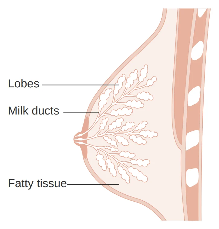 (full-size image, opens in a new tab)
Is DCIS cancer? Does it turn into invasive cancer?
DCIS is not invasive cancer, because the cells have not left the duct; the US National Cancer Institute also calls it a "precancer" [1]. But over time DCIS may grow through the duct wall and become invasive cancer [1]. How likely this is varies widely from person to person, and it is not yet possible to predict which DCIS will become invasive; that is why it is treated like cancer [1, 2].
Not all DCIS is alike. A type with a high grade and dead cells in the centre of the duct (comedo necrosis) appears more aggressive and is more likely to be found together with invasive cancer [2].
How is DCIS found? Calcifications on a mammogram
DCIS usually causes no symptoms and is most often found on a screening mammogram [1]. On a mammogram, DCIS often shows up as very small white specks called "calcifications" or "microcalcifications" [1]. DCIS rarely forms a lump that can be felt; occasionally it shows itself as a change in the breast or discharge from the nipple [1, 2].
If a mammogram shows something suspicious, a biopsy is done to find out whether it is DCIS, invasive cancer or something else [1]. Read about the numbers on a mammogram report in What does BI-RADS mean on a breast ultrasound or mammogram? and about biopsies in What is a biopsy and how long do the results take?
An important point: a needle biopsy removes only a small piece of the area. In an analysis of more than 7,000 patients, about 1 in every 4 whose needle biopsy showed DCIS was also found to have invasive cancer at surgery [4]. This is more likely with high-grade DCIS, an area larger than 2 cm, and when the DCIS looks like a lump on the mammogram [4]. That is why the final result is known only after surgery.
What does a DCIS pathology report say?
| On the report | What it means in plain language |
|---|---|
| Ductal carcinoma in situ (DCIS) | Abnormal cells inside a milk duct, not invading the surrounding tissue [1] |
| Nuclear grade: low, intermediate, high | Low, intermediate or high grade; a higher grade means the cells look more abnormal and the chance of recurrence is higher [1, 2] |
| Comedo necrosis: present | Dead cells are seen in the centre of the duct; a sign of a type that appears more aggressive [2] |
| Margins: negative (closest 3 mm) | No DCIS cells at the edges of the removed tissue; when radiotherapy follows surgery, a distance of 2 mm or more is enough [9] |
| ER: positive | Oestrogen receptor-positive; hormone therapy can lower the risk of a later cancer in the breast [10] |
| Microinvasion | Next to the DCIS, a very small invasive cancer, at most 1 mm, was also found; in that case the disease is no longer stage 0 [2] |
HER2 is not usually tested in DCIS [1]. For other terms, see How to read a pathology report and ER, PR, HER2 and Ki-67 on a breast cancer report: what do they mean?
How is DCIS treated? Surgery, radiotherapy and hormone therapy
- 1
Surgery
Surgery is almost always the first treatment [1]. Most often only the area of DCIS is removed with a layer of healthy tissue (breast-conserving surgery, or lumpectomy). If the DCIS is large or in several places in the breast, removal of the whole breast (mastectomy) may be recommended [1]. The surgeon sometimes also removes the nearest lymph node in the armpit [1]. If DCIS cells are found at the edges of the removed tissue, a second operation may be needed [9].
- 2
Radiotherapy
After breast-conserving surgery, the rest of the breast is usually treated with radiotherapy; after a mastectomy it is usually not needed [1]. In an analysis of 4 large studies with more than 3,700 women, radiotherapy cut the risk of the disease coming back in the same breast within 10 years by more than half, even in women with small, low-grade DCIS [5]. Over 10 years of follow-up, radiotherapy made no difference to the risk of dying of breast cancer [5]. For skin care, see Skin care during radiotherapy.
- 3
Hormone therapy, if the DCIS has hormone receptors
If the DCIS has oestrogen receptors, 5 years of tamoxifen tablets or, after the menopause, drugs such as anastrozole may be offered [1, 2]. In a large study, adding tamoxifen to surgery and radiotherapy cut the risk of a later invasive cancer in the same breast by about a third and also made cancer in the other breast less common [6]. Hormone therapy has not been shown to help people live longer, so the decision is made after talking through the benefits and side effects [2].
Can DCIS be managed without surgery? Active monitoring for low-risk DCIS
Researchers are studying whether low-risk DCIS can be watched with regular imaging instead of immediate surgery. In a US study of nearly 1,000 women aged 40 or older with grade 1 or 2, hormone receptor-positive DCIS, one group had imaging and an examination every 6 months and the other had the usual treatment [8]. After 2 years, invasive cancer in the same breast was not more common in the monitoring group, and in both groups it affected fewer than 6 in every 100 women [8].
These are short-term results and this approach is not yet standard treatment; the usual treatment for DCIS is still surgery [1, 8]. If you have low-risk DCIS and questions about this approach, talk to your surgeon and cancer specialist.
DCIS outlook: is it dangerous, and does it come back?
The outlook for DCIS is very good, and most people are cured with treatment [1]. More than 98 in every 100 people are alive 5 years after diagnosis [1]. In a study of more than 100,000 women in the United States, only about 3 in every 100 had died of breast cancer 20 years after a DCIS diagnosis; the risk was higher in women diagnosed before the age of 35 [7].
After treatment, the disease may come back in the same breast. About half of these recurrences are DCIS again and half are invasive cancer [6]. A recurrence as invasive cancer worsens the outlook, but a DCIS recurrence does not [6]. That is why regular follow-up with examinations and mammograms of both breasts matters. See Is breast cancer curable?
Carcinoma in situ in other organs: what does stage 0 mean?
"In situ" means cells that look like cancer cells under the microscope are found only in the place where they started and have not reached the surrounding tissue; this is also called "stage 0" [3]. These cells may one day become cancer and invade the healthy tissue around them [3]. Carcinoma in situ is also found in other organs [3]:
- Cervix, lungs and digestive tract: adenocarcinoma in situ. For the cervix, see What is a Pap smear? and What is HPV?
- Skin, mouth and larynx: squamous cell carcinoma in situ.
For the other stages, see What are the stages of cancer?
Frequently asked questions
Do I need to have my whole breast removed?
Do I need chemotherapy for DCIS?
Does DCIS spread to the lymph nodes or other parts of the body?
After DCIS, will I get cancer in the other breast too?
What is LCIS, and how is it different from DCIS?
Is DCIS hereditary? Do I need a genetic test?
If several people in your family have had breast or ovarian cancer, or you were diagnosed at a young age, ask your doctor about genetic counselling. See Genetic testing for cancer risk: who should be tested?
Related pages
Evidence on DCIS treatment and outcome (for doctors)
For physicians
Definition, stage and diagnosis. In the AJCC 8th edition DCIS = Tis (DCIS); LCIS is considered a benign entity and has been removed from TNM staging; T1mi = an invasive component of 1 mm or less. DCIS accounts for about 16% of all newly diagnosed invasive plus non-invasive breast tumours in the United States (2025 estimate) and about 25% of screen-detected cases, and more than 90% are diagnosed by mammography alone. The comedo type (high nuclear grade, pleomorphism and abundant central necrosis) is more aggressive [2]. Underestimation at needle biopsy (52 studies, 7,350 cases): 25.9% (95% CI 22.5 to 29.5); predictors: 14-gauge automated device versus 11-gauge vacuum-assisted biopsy, high grade, size over 20 mm on imaging, BI-RADS 4 or 5, a mass on mammography and palpability [4].
Radiotherapy and margins. EBCTCG 2010 (4 trials, 3,729 patients): 10-year absolute reduction in any ipsilateral breast event 15.2% (12.9% vs 28.1%), regardless of age, extent of surgery, tamoxifen, method of detection, margins, grade, comedonecrosis and size; even with negative margins and small low-grade lesions 18.0% (12.1% vs 30.1%); no significant effect on breast cancer mortality or all-cause mortality at 10 years [5]. RTOG 9804 (good-risk DCIS: mammographically detected, low or intermediate grade, under 2.5 cm, margins of 3 mm or more; 636 patients): 15-year in-breast recurrence 7.1% with radiotherapy vs 15.1% with observation (HR 0.36) [2]. Margins (SSO-ASTRO-ASCO 2016; 20 studies, 7,883 patients, with whole-breast irradiation): negative margins halve the risk of IBTR compared with ink on DCIS; 2 mm minimises the risk, and wider margins add no further benefit; a negative margin narrower than 2 mm is not on its own an indication for mastectomy [9].
Endocrine therapy. NSABP B-24 (1,804 patients; breast-conserving surgery and 50 Gy radiotherapy with tamoxifen 20 mg daily for 5 years or placebo): breast cancer events at 5 years 8.2% vs 13.4%, with no survival benefit [2]. Long-term follow-up of B-17 and B-24: 15-year I-IBTR 19.4% with lumpectomy alone, 8.9% with radiotherapy, 10.0% with radiotherapy plus placebo and 8.5% with radiotherapy plus tamoxifen; radiotherapy reduced I-IBTR by 52% (HR 0.48) and tamoxifen by 32% (HR 0.68); 53.7% of IBTRs were invasive, and I-IBTR was associated with higher mortality (HR 1.75) whereas DCIS recurrence was not; 15-year contralateral breast cancer 10.8% with placebo vs 7.3% with tamoxifen [6]. After the menopause: NSABP B-35 (3,104 patients) anastrozole versus tamoxifen gave fewer events (HR 0.73) without a survival benefit; IBIS-II DCIS (2,980 patients) showed no difference (HR 0.89) [2]. ER testing of DCIS is recommended and PgR is optional [10].
Outcome and active monitoring. SEER (108,196 women, 1988 to 2011): 20-year breast cancer-specific mortality 3.3%; with diagnosis before age 35, 7.8% vs 3.2% (HR 2.58); after an ipsilateral invasive recurrence, HR for death 18.1; after lumpectomy, radiotherapy lowered 10-year invasive recurrence (2.5% vs 4.9%) but not breast cancer-specific mortality (0.8% vs 0.9%) [7]. COMET (995 women aged 40 or older with grade 1 or 2, HR-positive DCIS without invasion): active monitoring every 6 months with imaging and examination versus guideline-concordant care (surgery ± radiotherapy); at a median follow-up of 36.9 months, the 2-year cumulative rate of ipsilateral invasive cancer was 4.2% vs 5.9% (difference −1.7%; upper limit of the 95% CI 0.95%), i.e. non-inferior; the primary outcome was assessed at 2 years [8].
Related cancer types
Related questions
References
11 referencesShow all referencesClose the reference list
Numbers in the text, such as [1], refer to these sources. Sources with a “Study card” button are summarised in the site’s evidence library.
- 1
- 2
- 3
- 4
- 5
- 6
- 7
- 8
- 9
- 10
- 11
This page is based on reputable scientific sources and was last updated on 28 October 2026. The content of this website is for educational and informational purposes only and does not replace consultation, diagnosis or treatment by a specialist physician. Consult your treating physician about any treatment decision.
Printed from the website; the full and up-to-date version: https://drdabiri.com/en/questions/dcis-stage-0/