A thyroid nodule is a small lump or growth in the thyroid gland. Nodules are very common: up to 6 in 10 adults have one, and only about 1 to 5 in every 100 nodules are cancer [2]. TI-RADS is a score that the radiologist gives from the appearance of the nodule on ultrasound; the higher the number, the higher the chance of cancer, but even at TI-RADS 4 most nodules are benign [1, 2]. Whether a biopsy is needed or ultrasound follow-up is enough depends on the TI-RADS level and the size of the nodule together [1].
Key points
- Thyroid nodules are very common, and most are not cancer [2, 6].
- TI-RADS runs from 1 to 5: 1 and 2 mean benign or not suspicious; 5 means the highest suspicion. But TI-RADS is not a diagnosis; it only shows which nodules need further tests [1, 2].
- Size matters too: in the American system, a TI-RADS 4 nodule is biopsied from 1.5 cm and a TI-RADS 5 nodule from 1 cm upwards; smaller nodules are followed up [1].
- The biopsy result is reported in 6 Bethesda categories: Bethesda 2 means benign and Bethesda 6 means cancer [4].
- "Indeterminate" results (Bethesda 3 and 4) are common, about 2 to 3 in every 10 biopsies; the next step is a repeat biopsy, a genetic test or surgery to make the diagnosis [5, 2].
- Even if a nodule is cancer, most thyroid cancers have a very good outcome with treatment [6, 7].
What is a thyroid nodule? Is a thyroid nodule dangerous?
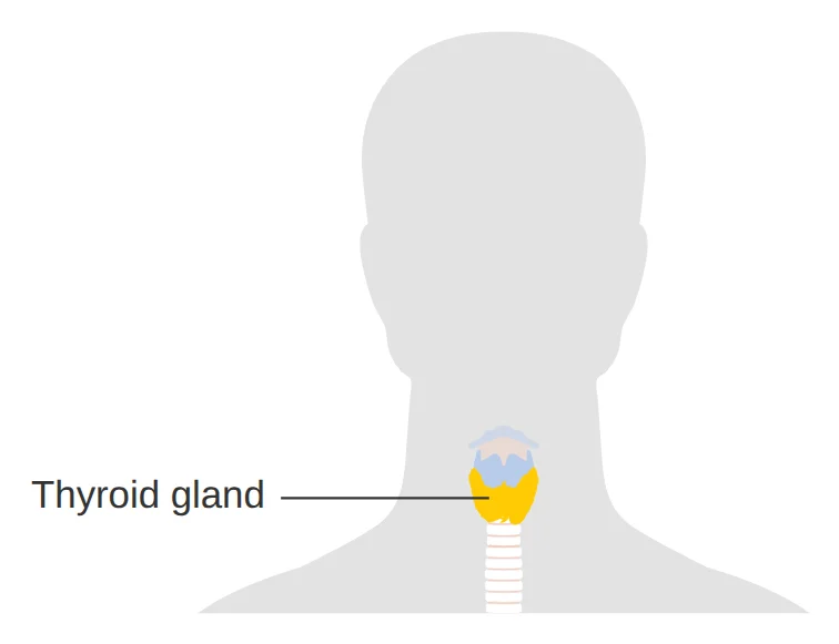
The thyroid is a butterfly-shaped gland in the front of the neck that makes thyroid hormone. A nodule is a lump or growth in this gland; it may be solid, filled with fluid (a cyst), or both [1].
Thyroid nodules are very common. Only a few are found by feeling the neck (about 5 in 100 women and 1 in 100 men), but ultrasound shows nodules in a large share of people [3]; up to 6 in 10 adults have one or more nodules [2]. Many nodules are found by chance, on scans done for other reasons [5].
Most nodules are not cancer. Of every 100 nodules found in the general population, usually only 1 to 5 are cancer [2]. This chance depends on age, sex, past radiation to the neck and family history [3]. A small number of nodules grow large enough to press on structures in the neck, or make extra hormone; each happens in about 1 in 20 nodules [5].
 (full-size image, opens in a new tab)
What is TI-RADS? Scoring a nodule on thyroid ultrasound
TI-RADS is a scoring method that the radiologist uses to estimate, from the ultrasound appearance of a nodule, how likely it is to be cancer, so that it is clear which nodules need a biopsy and which need only follow-up [1]. In the most widely used system (that of the American College of Radiology), five features of the nodule are assessed and each gets 0 to 3 points; the total sets the TI-RADS level [1]. The terms in brackets in the table below are the ones usually written in ultrasound reports.
| Feature | Lower risk | Higher risk |
|---|---|---|
| Make-up of the nodule (composition) | Cystic or almost completely cystic, or spongiform: 0 points [1] | Mixed cystic and solid: 1 point; solid or almost completely solid: 2 points [1] |
| How bright the nodule looks on the image (echogenicity) | Anechoic, like fluid: 0 points; same brightness as or brighter than thyroid tissue (isoechoic or hyperechoic): 1 point [1] | Darker than the thyroid (hypoechoic): 2 points; very dark (very hypoechoic): 3 points [1] |
| Shape | Wider than it is tall (wider-than-tall): 0 points [1] | Taller than it is wide (taller-than-wide): 3 points [1] |
| Edge (margin) | Smooth or ill-defined: 0 points [1] | Lobulated or irregular: 2 points; spreading outside the thyroid (extrathyroidal extension): 3 points [1] |
| Bright spots, including calcifications (echogenic foci) | None, or large comet-tail artefacts: 0 points [1] | Macrocalcifications: 1; calcification around the edge of the nodule (rim): 2; tiny bright spots (punctate echogenic foci): 3 points [1] |
One option is chosen from each row, except for echogenic foci, where the points of all types present are added together [1].
TI-RADS 3, TI-RADS 4 and TI-RADS 5: what do they mean and when does a suspicious nodule need a biopsy?
| Level | Total points | Meaning | Biopsy if the nodule is | Ultrasound follow-up if the nodule is |
|---|---|---|---|---|
| TI-RADS 1 (TR1) | 0 | Benign | Not needed | Not needed [1] |
| TI-RADS 2 (TR2) | 2 | Not suspicious | Not needed | Not needed [1] |
| TI-RADS 3 (TR3) | 3 | Mildly suspicious | 2.5 cm or larger | 1.5 cm or larger [1] |
| TI-RADS 4 (TR4) | 4 to 6 | Moderately suspicious | 1.5 cm or larger | 1 cm or larger [1] |
| TI-RADS 5 (TR5) | 7 or more | Highly suspicious | 1 cm or larger | 0.5 cm or larger [1] |
The European guideline and the American Thyroid Association use slightly different sizes [2, 3]. Your doctor makes the final decision, taking your situation into account.
What do TI-RADS 1 and TI-RADS 2 mean? The nodule is benign or not suspicious and does not need a biopsy [1]. For example, a simple cyst (filled with fluid) scores zero points [1].
What does TI-RADS 3 mean? The nodule is mildly suspicious. In the European system, only 2 to 4 in every 100 nodules in this category are cancer [2]. A biopsy is needed only for large nodules (2.5 cm or more in the American system) [1].
What does TI-RADS 4 mean? The nodule is moderately suspicious. In the European system, 6 to 17 in every 100 nodules in this category are cancer; in other words, most TI-RADS 4 nodules are also benign [2]. In the American system, a biopsy is done from 1.5 cm upwards [1]; a smaller nodule is followed up with ultrasound, and the European guideline suggests a repeat ultrasound after one year [2].
What does TI-RADS 5 mean? The nodule is highly suspicious. In the European system, about 3 to 9 in every 10 nodules in this category are cancer [2]. A biopsy is done from 1 cm upwards [1]. The European guideline follows a smaller nodule with ultrasound every 6 to 12 months, unless a lymph node in the neck looks suspicious, the nodule may have grown outside the thyroid, or it lies close to the windpipe or the voice nerve; in that case a biopsy is advised [2].
There are several TI-RADS systems, such as the American and the European systems, and none has always been better than the others [2]. The numbers do not mean exactly the same thing in every system, and sometimes category 4 is written with a letter (such as 4a or 4b). So read your ultrasound result together with your own doctor.
Thyroid nodule with calcification: is it dangerous?
Calcification means deposits of calcium, which show up on ultrasound as bright spots. The type matters [1]:
- Tiny bright spots ("microcalcifications" or punctate echogenic foci in reports): these score the most points (3 points) and in the European system are one of the features of high suspicion [1, 2].
- Calcification around the edge of the nodule (rim or peripheral): 2 points [1].
- Coarse calcification (macrocalcification): 1 point [1].
Calcification on its own does not mean cancer; its points are added to those of the nodule's other features to give the TI-RADS level [1].
Needle biopsy (FNA) and Bethesda results 1 to 6
First of all, the doctor orders a TSH blood test (the hormone that stimulates the thyroid) [2, 3]. If TSH is low, a thyroid scan is done to see whether the nodule is "hot" (overactive); hot nodules are rarely cancer and are not biopsied [2].
If a biopsy is needed, a thin needle is passed through the skin into the thyroid to remove a little tissue from the nodule [6]. A pathologist reports the result in one of the 6 Bethesda categories, and each category carries a known chance of cancer [4].
| Category | Name | Chance of cancer | Usual next step |
|---|---|---|---|
| Bethesda 1 | Nondiagnostic | About 1 in 8 [4] | Repeat biopsy [2, 4] |
| Bethesda 2 | Benign | About 1 in 25 [4] | Ultrasound follow-up; in the European guideline usually after 3 to 5 years [2, 4] |
| Bethesda 3 | Atypia of undetermined significance (AUS) | About 1 in 5 [4] | Repeat biopsy; sometimes a genetic test, follow-up or surgery [2, 4] |
| Bethesda 4 | Follicular neoplasm | About 1 in 3 [4] | Usually surgery to make the diagnosis (removal of half of the thyroid); sometimes a genetic test [2, 4] |
| Bethesda 5 | Suspicious for cancer | About 3 in 4 [4] | Usually surgery [2, 4] |
| Bethesda 6 | Malignant (cancer) | Almost all [4] | Surgery; for some very small cancers, regular monitoring [2, 4] |
The decision is always made taking the ultrasound, the size of the nodule and your situation into account.
Bethesda 3 and 4: what does an "indeterminate" result mean and what happens next?
About 2 to 3 in every 10 thyroid biopsies give an "indeterminate" result [5]; this means the cells are neither completely normal nor clearly cancer. Most of these nodules are not cancer either: about 1 in 5 people with Bethesda 3 and about 1 in 3 with Bethesda 4 have cancer [4].
- Repeat biopsy: for Bethesda 3, the European guideline suggests repeating the biopsy [2].
- A genetic (molecular) test on the sample: helps the decision when the result is indeterminate [5, 2].
- Regular monitoring: if a Bethesda 3 result comes back again, regular monitoring is one of the options [2].
- Surgery to make the diagnosis: removal of half of the thyroid so that the whole nodule can be examined under the microscope; for Bethesda 4, surgery is usually offered [2, 4].
When is surgery needed, and when is follow-up enough?
- Benign nodule (Bethesda 2): usually ultrasound follow-up only; the European guideline suggests an ultrasound after 3 to 5 years, but if the nodule looks highly suspicious on ultrasound (TI-RADS 5), the biopsy is repeated [2].
- A benign nodule that presses on structures in the neck: surgery, or heat treatment through the skin (destroying the nodule with a needle) instead of surgery [2].
- A nodule that grows during follow-up: the American Thyroid Association counts a nodule as having grown if at least two of its measurements increase by a fifth (and by at least 2 mm), or its volume increases by more than half [3]. In that case your doctor decides whether to repeat the biopsy.
- Suspicious for cancer or cancer (Bethesda 5 and 6): usually surgery. For nodules of 5 to 10 mm without suspicious lymph nodes and with no risk of growth outside the thyroid, regular monitoring or minimally invasive treatments (such as heat treatment through the skin) are also possible [2]. On monitoring very small cancers, see Active surveillance for papillary microcarcinoma.
If the biopsy shows cancer, know that most thyroid cancers can be cured [6]: Is thyroid cancer curable?
When should I see a doctor?
Most thyroid nodules cause no symptoms. Tell your doctor promptly about these:
- A new lump in the neck, or a nodule that is getting bigger [6, 3]
- Hoarseness that does not go away [6]
- Difficulty or pain when swallowing [6]
- Difficulty breathing [6]
If you had radiation to the head and neck as a child or thyroid cancer runs in your family, tell your doctor this too, because the chance of cancer in a nodule is higher [3, 6].
Severe shortness of breath or being unable to swallow needs urgent care; call your local emergency number.
Frequently asked questions
What does a "malignant" thyroid nodule mean?
Malignant means cancer, and benign means not cancer. Most thyroid nodules are benign [2]. Even if a nodule is cancer, most thyroid cancers have a very good outcome with treatment [7]. See Is thyroid cancer curable? and What is the difference between a benign and a malignant tumour?
My small nodule is TI-RADS 5 (for example 6 mm); does it need a biopsy?
Usually not. In the American system, a TI-RADS 5 nodule is biopsied from 1 cm upwards and followed up from 0.5 cm upwards [1]. For a highly suspicious nodule smaller than 1 cm, the European guideline suggests an ultrasound every 6 to 12 months and advises a biopsy when a lymph node in the neck looks suspicious, the nodule may have grown outside the thyroid, or it lies close to the windpipe or the voice nerve [2].
Is a thyroid cyst dangerous?
Do I need a thyroid ultrasound as a check-up?
If you have no symptoms, no; the US Preventive Services Task Force recommends against screening for thyroid cancer in adults without symptoms, because the harms of finding and treating harmless cancers outweigh the benefit [7]. If you feel a lump in your neck or have one of the symptoms above, see a doctor.
What is a "hot" nodule?
A hot (overactive) nodule makes thyroid hormone itself and shows up more strongly than the rest of the gland on a thyroid scan. When the TSH level in the blood is low, a scan is done to find these nodules. Hot nodules are rarely cancer and are not biopsied [2].
Does Bethesda 3 mean I have cancer?
Can a thyroid biopsy make cancer spread?
A thin-needle biopsy is the main way to diagnose a suspicious nodule [6, 2]. Read more about this worry: Does a biopsy make cancer spread?
TI-RADS, Bethesda and decision-making criteria (for physicians)
For physicians
ACR TI-RADS (2017). Points: composition (cystic or spongiform 0, mixed 1, solid 2), echogenicity (anechoic 0, hyper- or isoechoic 1, hypoechoic 2, very hypoechoic 3), shape (wider-than-tall 0, taller-than-wide 3), margin (smooth or ill-defined 0, lobulated or irregular 2, extrathyroidal extension 3) and echogenic foci (none or large comet-tail 0, macrocalcifications 1, peripheral calcifications 2, punctate echogenic foci 3; all present are added). TR1 (0 points) and TR2 (2 points): no FNA; TR3 (3): FNA from 2.5 cm and follow-up from 1.5 cm; TR4 (4 to 6): FNA from 1.5 cm and follow-up from 1 cm; TR5 (7 or more): FNA from 1 cm and follow-up from 0.5 cm [1].
EU-TIRADS (ETA 2023). Risk of malignancy: EU-TIRADS 2 0%; 3 (iso- or hyperechoic without high-suspicion features) 2% to 4%; 4 (mildly hypoechoic without high-suspicion features) 6% to 17%; 5 (at least one of: irregular shape, irregular margins, microcalcifications, marked hypoechogenicity) 26% to 87%. FNA thresholds: more than 20, 15 and 10 mm for categories 3, 4 and 5. Follow-up of nodules not biopsied: EU-TIRADS 2 over 10 mm and EU-TIRADS 3 between 10 and 20 mm every 3 to 5 years; 5 to 10 mm, no further evaluation; EU-TIRADS 4 under 15 mm after 1 year; EU-TIRADS 5 under 10 mm every 6 to 12 months. For EU-TIRADS 5 nodules of 5 to 10 mm, FNA is recommended if there are suspicious lymph nodes, a risk of extrathyroidal extension or a worrisome location (e.g. close to the trachea or the recurrent laryngeal nerve). No system has been consistently superior, and in Europe EU-TIRADS is preferred [2].
ATA 2015. Nodule prevalence 19% to 68% on high-resolution ultrasound and about 5% in women and 1% in men by palpation; cancer in 7% to 15% of nodules depending on age, sex, radiation history and family history. Patterns: high suspicion (risk >70% to 90%; FNA from 1 cm), intermediate suspicion (10% to 20%; from 1 cm), low suspicion (5% to 10%; from 1.5 cm), very low suspicion (<3%; from 2 cm or observation), benign (<1%; no FNA). Measure TSH at the initial evaluation and perform radionuclide scanning if TSH is low. Growth: a 20% increase in at least two dimensions with a minimum of 2 mm, or more than 50% in volume [3].
Bethesda 2023 (mean risk of malignancy and range). I nondiagnostic 13% (5 to 20); II benign 4% (2 to 7); III AUS 22% (13 to 30); IV follicular neoplasm 30% (23 to 34); V suspicious for malignancy 74% (67 to 83); VI malignant 97% (97 to 100). In the third edition, each category has a single name, and AUS is simplified into two subgroups based on risk and molecular profile [4]. Indeterminate cytology accounts for about 20% to 30% of all FNAs, and molecular testing is indicated only in selected cases, such as indeterminate cytology [5].
ETA 2023 recommendations by cytology. Bethesda I: repeat FNA; if still nondiagnostic, core needle biopsy (CNB), or molecular testing in EU-TIRADS 4 and 5; then follow-up or surgery. Bethesda II: in EU-TIRADS 3 and 4, re-evaluation after 3 to 5 years, and in EU-TIRADS 5, repeat FNA. Bethesda III: repeat FNA regardless of EU-TIRADS category; if Bethesda III again, re-evaluation within 1 year, molecular testing or surgery for EU-TIRADS 3, and surgery or active surveillance (with molecular testing if available) for EU-TIRADS 4 and 5. Bethesda IV: offer surgery and molecular testing. Bethesda V and VI: recommend surgery; for 5 to 10 mm nodules without suspicious lymph nodes or risk of extrathyroidal extension, active surveillance or minimally invasive treatment (MIT) may be considered. Hyperfunctioning nodules are rarely malignant and are not biopsied; scintigraphy when TSH is low. Consider calcitonin for nodules scheduled for surgery or MIT and for indeterminate cytology or suspicious ultrasound features, and measure it with a personal or family history of medullary cancer or MEN2 (cut-offs >30 pg/mL in women and >34 pg/mL in men). Thermal ablation (TA) is an alternative to surgery for symptomatic solid benign nodules [2].
Screening. The USPSTF (2017, grade D) recommends against screening for thyroid cancer in asymptomatic adults [7].
Related cancer types
Related questions
References
7 referencesShow all references, 4 with a study cardClose the reference list
Numbers in the text, such as [1], refer to these sources. Sources with a “Study card” button are summarised in the site’s evidence library.
- 1
- 2
- 3
- 4
- 5
- 6
- 7
This page is based on reputable scientific sources and was last updated on 28 October 2026. The content of this website is for educational and informational purposes only and does not replace consultation, diagnosis or treatment by a specialist physician. Consult your treating physician about any treatment decision.
Printed from the website; the full and up-to-date version: https://drdabiri.com/en/questions/thyroid-nodule-tirads/