A PET scan is a type of imaging that uses an injection of a small amount of radioactive material — usually labelled sugar (FDG) — to show the metabolic activity of tissues, and today it is usually done together with a CT scan (PET-CT) [1, 2]. In cancer it is used to tell benign from malignant lesions, for staging, to assess response to treatment, to find recurrence, to choose a biopsy site and to plan radiotherapy [1]. FDG is also taken up by infection, inflammation and healing tissue; so not every bright spot means cancer [1, 2].
Examples of how PET scans are used in cancer
- Lymphoma: to see how far the disease has spread and how well treatment has worked [3].
- High-risk prostate cancer: a special type of PET (PSMA) shows the spread of disease more accurately than CT and a bone scan [4].
- Oesophageal cancer: before surgery, it sometimes shows spread that was not seen on other imaging [5].
- Brain: a standard PET does not help much for the brain, and MRI is usually better [1].
For physicians
| Situation | Use | Note |
|---|---|---|
| Lymphoma | Standard staging and response assessment (with a 5-point scale) in FDG-avid types [3] | In Hodgkin lymphoma and most diffuse large B-cell lymphomas, a bone marrow biopsy is no longer routinely needed for staging [3] |
| High-risk prostate cancer | Staging with PSMA-based PET-CT [4] | More accurate than CT and bone scan (accuracy 92% vs 65%) [4] |
| Oesophageal cancer | Looking for occult metastases before surgery [5] | Shows occult metastases in about 15% of surgical candidates [5] |
| Brain metastases | Limited value with FDG, because of the high normal uptake of FDG in the brain [1] | MRI is usually preferred |
Preparation and the day of the test
- Fasting: have nothing but plain water for at least 4 hours before the test [1].
- Enough water: drinking about one litre of water in the 2 hours before the injection is recommended [1].
- Do not exercise: avoid strenuous activity for at least 6 hours, and preferably 24 hours, before the test [1].
- Diabetes: blood sugar is measured before the injection; if you have diabetes or use insulin, arrange this with the imaging centre in advance [1].
- Pregnancy and breastfeeding: always mention a possible pregnancy; breastfeeding mothers should limit close contact with their baby for 12 hours after the injection [1].
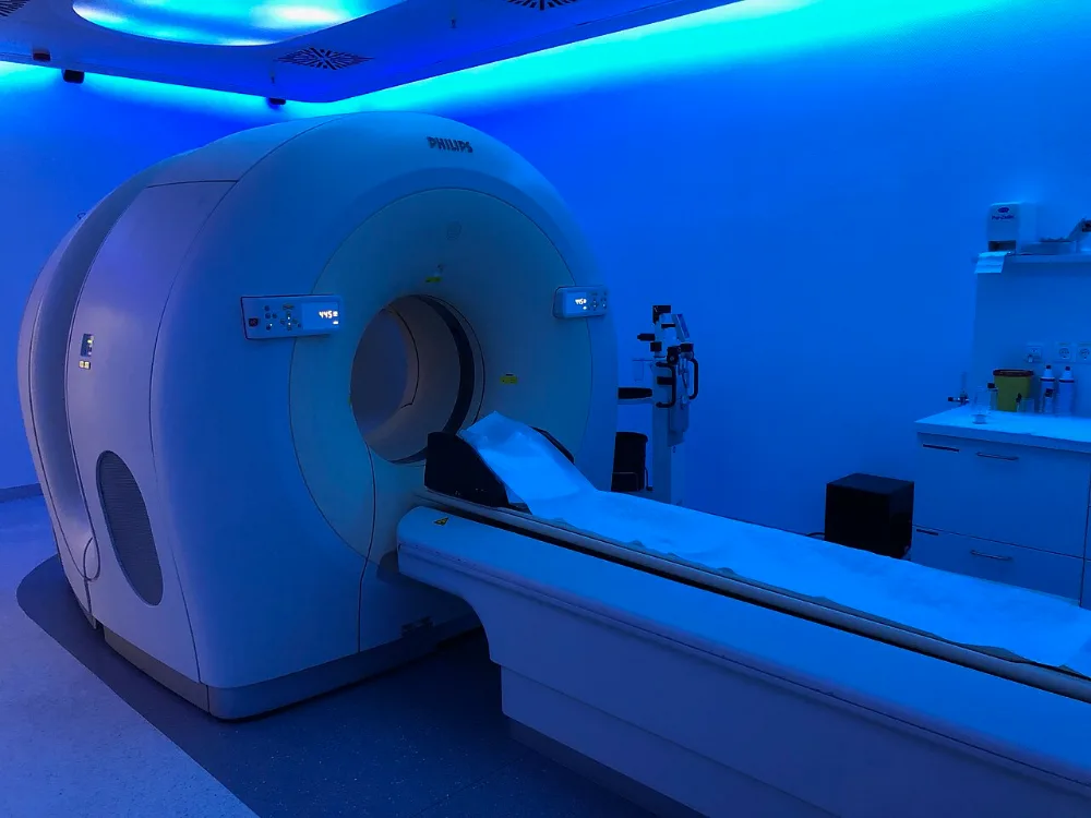
What happens on the day of the test?
- 1
Measuring blood sugar
Before the FDG injection, blood sugar is measured [1].
- 2
Injection and rest
After the injection, you rest for about 60 minutes sitting or lying down, somewhere warm and quiet, while the substance spreads through your body [1].
- 3
Imaging
You lie on the scanner bed and PET and CT images are taken.
- 4
Report
A nuclear medicine specialist interprets the images together with the CT findings and your history; combining PET with CT makes interpretation more accurate [2].
When should I call my doctor?
If after the test you have severe swelling, pain or redness at the injection site, contact the centre or your doctor.
Related questions
They saw a bright spot on my PET scan; does that mean the cancer has spread?
Not necessarily. Inflammation, infection, injuries after trauma or surgery and benign tumours also take up FDG [2]. In one review, more than 1 in 4 cancer patients had a bright spot on the PET scan that was not cancer; most were due to inflammation [2]. Sometimes a biopsy or other imaging is needed to be sure.
Why do I need to wait for a PET scan after chemotherapy or surgery?
So that inflammatory changes caused by treatment do not cause errors; the EANM guideline gives an interval of at least 10 days after chemotherapy, 2 to 3 months after radiotherapy and at least 6 weeks after surgery (to assess the operation site) [1].
Does a PET scan replace a biopsy?
Usually not; one use of a PET scan is to choose the best site for a biopsy [1]. In most cases, the firm diagnosis of cancer is made with a biopsy.
Related cancer types
References
5 referencesShow all references, 5 with a study cardClose the reference list
Numbers in the text, such as [1], refer to these sources. Sources with a “Study card” button are summarised in the site’s evidence library.
- 1
- 2
- 3
- 4
- 5
This page is based on reputable scientific sources and was last updated on 28 October 2026. The content of this website is for educational and informational purposes only and does not replace consultation, diagnosis or treatment by a specialist physician. Consult your treating physician about any treatment decision.
Printed from the website; the full and up-to-date version: https://drdabiri.com/en/questions/what-is-a-pet-scan/