A cancerous mole (melanoma) is usually a mole that looks different from your other moles or is changing: its two halves do not match, its edge is irregular, it has several colours, it is usually larger than 6 mm, or its size, shape or colour changes and it starts to itch or bleed [1, 2, 3]. Some melanomas do not look like this: a firm lump that keeps growing, sometimes pink or red, a brown or black streak under a nail, or a dark spot on the sole of the foot or the palm [3, 4, 5]. Most moles are harmless, but show any new or changing mole to a doctor.
Key points
- Most moles are harmless. Most adults have 10 to 40 moles, and most melanomas do not start in a mole you already had [2].
- Change is often the first sign: a mole whose size, shape or colour changes, or that starts to itch, hurt or bleed [1, 2].
- A mole that looks different from the others (the "ugly duckling") and a firm lump that keeps growing should also be shown to a doctor promptly, even if they do not fit the ABCDE rule [3].
- Melanoma can appear under a nail or on the sole of the foot. In a study from Tehran, Iran, this type on the palms, soles and nails was the most common type of melanoma [7].
- A painless mole is not necessarily a harmless one; melanomas usually neither hurt nor itch [8].
- Removing a suspicious mole for testing is the main way to make the diagnosis, and in a large study, taking a sample before the full operation did not worsen the outcome [9, 10].
What does a normal mole look like?
A normal mole is usually smaller than 5 mm (the width of a pencil eraser), round or oval, with a smooth surface and a clear edge, and is often slightly raised. Its colour is even: pink, tan or brown [2].
Most adults have 10 to 40 moles. Most moles are above the waist, in places that get sun. Most people keep getting new moles until about the age of 40, and moles tend to fade in older age [2].
Some moles are "atypical" (dysplastic): larger than 5 mm and with a different shape or colour. These moles also rarely turn into cancer, but people who have many of them are at higher risk of melanoma [2].
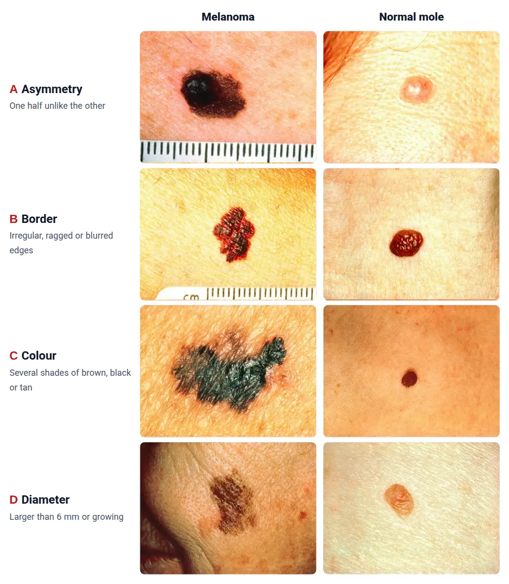
What does a cancerous mole look like? The ABCDE rule
Doctors use a simple rule called ABCDE to recognise a suspicious mole [1, 2]:
- A, asymmetry: the two halves of the mole do not match [2].
- B, border: the edges are ragged, notched or blurred [2].
- C, colour: several shades of black, brown and tan in one mole; sometimes areas of white, grey, red, pink or blue [2].
- D, diameter: most melanomas are larger than 6 mm and grow, but they can also be very small [2].
- E, evolving: the mole has changed over the past few weeks or months: in size, shape, colour, surface (for example, bleeding) or feel (itching or tenderness) [1, 2].
Having one of these signs does not mean the mole is cancer; it means a doctor should look at it. Doctors place special weight on change [1].
How to know if a mole is cancerous: the mole that looks different
A person's moles usually look alike. A mole that looks different from your other moles, for example darker, larger or a different shape, should be checked. Doctors call this the "ugly duckling" sign [3]. It finds melanomas as well as the ABCDE rule does and flags fewer harmless moles as suspicious [3].
To notice changes, check all your skin from time to time in good light and with a mirror, including the soles of your feet, between your toes, your back and your scalp. People with atypical moles should check their skin once a month [2]. Take a photo of a mole that worries you, with a ruler next to it, so that you can compare later.
What does melanoma look like when it is a raised, fast-growing lump? Nodular and colourless melanoma
Some melanomas do not show the ABCDE signs. One of these is nodular melanoma (a raised melanoma). Doctors remember three signs for it [3]:
- Elevated: a new raised spot or thickening of the skin
- Firm: it feels firm to the touch
- Growing: it is getting bigger
Some melanomas are also colourless (amelanotic): skin-coloured, pink or red. For these, the simple sign is red, raised and recently changed [3]. Do not wait for such a lump to go away on its own; show it to a doctor promptly.
Melanoma under the nail: is a dark streak a bruise or cancer?
Nail melanoma is rare and is often diagnosed late [5]. About 2 in 3 nail melanomas start as a brown or black streak along the length of the nail [5]. These signs make cancer more likely [4, 5]:
- A wide streak (3 mm or more), several colours, or a streak that gets darker or wider; especially if the base of the streak (near the skin) is wider than its tip [4, 5]
- Dark colour spreading onto the skin beside or at the base of the nail [4]
- A streak on the thumb or the big toe, the most common sites of nail melanoma [5]
- A split nail, bleeding, or a pink or red lump under the nail; some nail melanomas are not dark [5]
- A change in the nail that does not improve with treatment, for example treatment for a fungal infection [4, 3]
A bruise under the nail (a collection of blood), for example after an injury, is often purple to black, and the key difference is that it gradually moves towards the tip of the nail as the nail grows [6]. If a mark does not move forward as the nail grows, or it is a line that starts at the base of the nail and runs to the tip, show it to a doctor [6, 5]. A dark nail streak has many different causes and is not always cancer; the diagnosis is confirmed by a biopsy of the nail root [5].
Melanoma on the sole of the foot and the palm
One type of melanoma appears on the soles of the feet, the palms and under the nails. It is rare in white people, but in people with darker skin and in East Asia, a large share of melanomas are of this type [3]. In a study of 450 patients in Tehran, Iran, this type on the palms, soles and nails was also the most common type of melanoma (about 3 in 10 patients), and nearly half of the tumours were on the arms and legs [7].
This type of melanoma is sometimes mistaken for other things, such as a wart, a diabetic foot ulcer or a fungal nail infection [3]. So show these to a doctor:
- A new brown or black spot on the sole of the foot or the palm, or a spot that becomes larger, more irregular or multicoloured [1, 3]
- A "wart" or sore on the sole of the foot that has not healed within 2 months despite treatment or is getting worse, especially if it bleeds [3, 13]
Remember the soles of your feet and between your toes when you check your skin.
Is skin cancer itchy? Does a cancerous mole hurt?
Usually not. In a study of more than 300 skin cancers, about 4 in 10 itched and about 3 in 10 were painful; but this was mostly in skin cancers other than melanoma, and melanomas mostly had neither pain nor itching [8]. So a painless mole is not necessarily a harmless one.
On the other hand, if a mole that has stayed the same for years starts to itch, feel tender, ooze or bleed, that is also a kind of change and should be checked [1, 2]. What matters is new itching or bleeding in a mole.
When should I see a doctor?
If you have any of these, see a family doctor or a dermatologist and do not wait for it to get better on its own:
- A new mole that looks different from your other moles, or a mole whose size, shape or colour has changed over a few weeks or months [1, 2, 3]
- A mole that has started to itch, bleed, ooze or form a sore [1, 2]
- A firm lump, pink, red or dark, that is getting bigger [3]
- A new brown or black streak in a nail, especially if it is getting wider or darker or its colour has spread to the skin around the nail [4, 5]
- A dark spot on the sole of the foot or the palm that is changing, or a sore on the foot that does not heal [3, 13]
If a mole is bleeding or growing quickly, do not put off seeing a doctor. Most of these changes have a harmless cause, but only a doctor can tell.
What happens at the doctor? Does removing a mole spread cancer?
- 1
Skin examination
The doctor looks at the mole closely, often with a special lighted skin magnifier (dermatoscope), and compares it with your other moles [3].
- 2
- 3
Examination under the microscope
A pathologist examines the sample and determines whether the mole is cancer. To lower the chance of a mistake, a second review of the sample by another pathologist can also be considered [9].
Some people fear that touching a mole or taking a sample will spread cancer. In a study of more than 700 patients in Scotland, people who first had only part of their melanoma removed for testing were compared with similar patients who had the whole mole removed from the start. The type of biopsy did not increase the chance of the disease coming back or of death from melanoma [10]. So fear of a biopsy should not delay the diagnosis; read more: Does a biopsy make cancer spread?
A suspicious mole should not be shaved off or burned off; it is best removed whole and examined under the microscope [9]. If you want a mole removed for cosmetic reasons, show it to a doctor first.
If a cancerous mole is found early, can it be cured?
Most of the time, yes. In a large study in the United States, for the most common type of melanoma (the type that spreads across the surface of the skin), more than 9 in 10 patients were alive 10 years later in the final years of the study [11]. In the same study, raised (nodular) melanoma made up about 1 in 7 melanomas but accounted for more than a third of deaths from melanoma [11]; that is why a firm lump that keeps growing should be shown to a doctor promptly.
In the Tehran study, nearly 1 in 4 patients were diagnosed when the tumour had already become thick [7]. That is why showing a suspicious mole to a doctor early matters. For melanoma treatment, see Melanoma and Treatment of advanced melanoma.
Frequently asked questions
What does "malignant" mean for a mole? Can a benign mole turn into cancer?
Melanoma is a cancer, and every cancer is "malignant"; a normal mole is "benign", which means it is not cancer. A normal mole, and even an atypical (dysplastic) mole, rarely turns into melanoma, and most melanomas do not start in an existing mole [2]. See the difference in What is the difference between a benign and a malignant tumour?
I have a lot of moles; should I have them all removed?
No. Normal moles, or even atypical moles, usually do not need to be removed [2]. People with atypical moles should check their skin once a month and have their skin examined by a doctor regularly [2]. Show a doctor any mole that is changing or looks different from the others, so that they can decide whether it should be removed [1, 3].
I have a new mole after the age of 40; should I worry?
Skin cancer pictures: can you tell from a photo whether a mole is cancerous?
Pictures help you learn the warning signs, but the look of a mole alone is not enough; a doctor examines the mole and, if it is suspicious, removes it for examination under the microscope [9]. Taking photos of your own mole at regular intervals is useful for seeing whether it changes.
Is a mole on the sole of the foot dangerous?
Not every mole on the sole is cancer. But melanoma on the sole is sometimes mistaken for a wart or a sore and is diagnosed late [3]. Show a doctor any new or changing dark spot on the sole of the foot or the palm.
I have a black line on my nail; is it cancer?
Not necessarily; a dark nail streak has many different causes [5]. But a new streak in one nail, a wide or multicoloured streak, a streak that is getting wider or darker, or colour that has spread to the skin around the nail should be checked [4, 5]. See the section Melanoma under the nail.
What does skin cancer look like? Is it always a mole?
No. Melanoma is only one type of skin cancer. The more common skin cancers (basal cell and squamous cell carcinoma) look different; see their signs in What are the symptoms of skin cancer? and Skin cancer.
Clinical criteria and diagnostic points (for physicians)
For physicians
ABCDE. Abbasi 2004 (JAMA, review of evidence from 1980 to 2004): the data did not support lowering the diameter criterion (more than 6 mm) but supported adding E (evolving): changes in size, shape, symptoms (itching, tenderness), surface (especially bleeding) and shades of colour [1]. The "ugly duckling" sign (a lesion that differs from the patient's other naevi) is more specific than ABCDE with similar sensitivity; the EFG rule (Elevation, Firmness, Growth) has been proposed for nodular melanoma, the 3R rule (Red, Raised, Recent change) for amelanotic melanoma, and the CUBED and ABCDEF rules for acral and subungual melanoma [3].
Weighted 7-point checklist (7PCL). Major criteria (2 points each): change in size, irregular pigmentation, irregular border; minor criteria (1 point each): inflammation, itch or altered sensation, larger than other lesions (diameter >7 mm), oozing or crusting. The NICE recommendation (2005) was referral for a score of 3 or more. In the Walter 2013 validation in primary care, a cut-off of 4 gave a sensitivity for melanoma of 91.7% and a specificity of 53.4% (for clinically significant lesions: sensitivity 73.3%, specificity 57.1%, positive predictive value 24.1% and negative predictive value 92.0%) [12].
Nail unit melanoma. 0.7% to 3.5% of melanomas; Levit's ABCDEF: A, age (peak in the fifth to seventh decades) and race (up to one third of melanomas in African Americans, Asians and Native Americans); B, a brown to black band 3 mm or more wide with variegated borders; C, change in the band or no improvement of the nail dystrophy despite adequate treatment; D, the digit involved; E, extension of pigment onto the nail fold (Hutchinson's sign); F, family or personal history of dysplastic naevi or melanoma [4]. About two thirds present as longitudinal melanonychia; a width of more than 3 mm or more than 40% of the nail plate, colour heterogeneity, darkening or widening, and a triangular band (wider proximally) are warning features; up to 25% are amelanotic. 58% of cases on the fingers involve the thumb and 86% of cases on the toes involve the hallux; periungual pigment alone is not diagnostic, and matrix biopsy is the gold standard [5]. In 47 subungual haematomas, the diagnosis was confirmed by distal migration of the colour change; a blue-white colour (a melanoma clue) was seen in 26%, so follow-up until the colour reaches the free edge of the nail is advised [6].
Acral melanoma. It makes up the largest share of melanomas in East Asian (50% to 58%), Hispanic (49% to 61%) and Black (39% to 75%) populations; the misdiagnosis rate is 34%, and common misdiagnoses are warts, diabetic foot ulcers and onychomycosis [3]. In 450 patients at a referral centre in Tehran (2004 to 2014): acral lentiginous 30% and lentigo maligna 29%; 49% on the extremities; mean Breslow thickness 2.8 mm, 38% less than 1 mm and 23% more than 4 mm [7].
Pain and itch. In 339 skin cancers (166 BCC, 146 SCC, 27 melanomas), the prevalence of itch was 36.9% and of pain 28.2%, but these were mostly absent in melanomas; pain intensity was associated with inflammation, ulceration, perineural invasion and depth of invasion [8].
Biopsy. Biopsy preferably by complete local excision; a suspicious lesion should never be shaved off or cauterised; consider a second review by an independent pathologist [9]. In a case–control study from the Scottish Melanoma Group, 265 incisional biopsies were compared with 496 excisional biopsies (matched for age, sex, site and Breslow thickness); biopsy type had no effect on recurrence (P=0.30) or melanoma-related death (P=0.34) [10].
Nodular subtype and melanoma deaths. Shaikh 2012 (original 9 SEER registries, 1978 to 2007, 111,478 patients): superficial spreading melanoma made up 66% of cases and 46% of ultimately fatal melanomas, and nodular melanoma 14% of cases and 37% of ultimately fatal melanomas; 10-year relative survival for superficial spreading melanoma rose from 90.6% to 96.5%, but for nodular melanoma it remained stable (61.8% to 61.5%) [11]. These data show the importance of teaching the EFG rule.
Related cancer types
References
13 referencesShow all referencesClose the reference list
Numbers in the text, such as [1], refer to these sources. Sources with a “Study card” button are summarised in the site’s evidence library.
- 1
- 2
- 3
- 4
- 5
- 6
- 7
- 8
- 9
- 10
- 11
- 12Using the 7-point checklist as a diagnostic aid for pigmented skin lesions in general practice: a diagnostic validation study
- 13
This page is based on reputable scientific sources and was last updated on 28 October 2026. The content of this website is for educational and informational purposes only and does not replace consultation, diagnosis or treatment by a specialist physician. Consult your treating physician about any treatment decision.
Printed from the website; the full and up-to-date version: https://drdabiri.com/en/questions/cancerous-mole/